Saturday 18 July 2020

BiPaddle Pectoralis major musculocutaneous flap


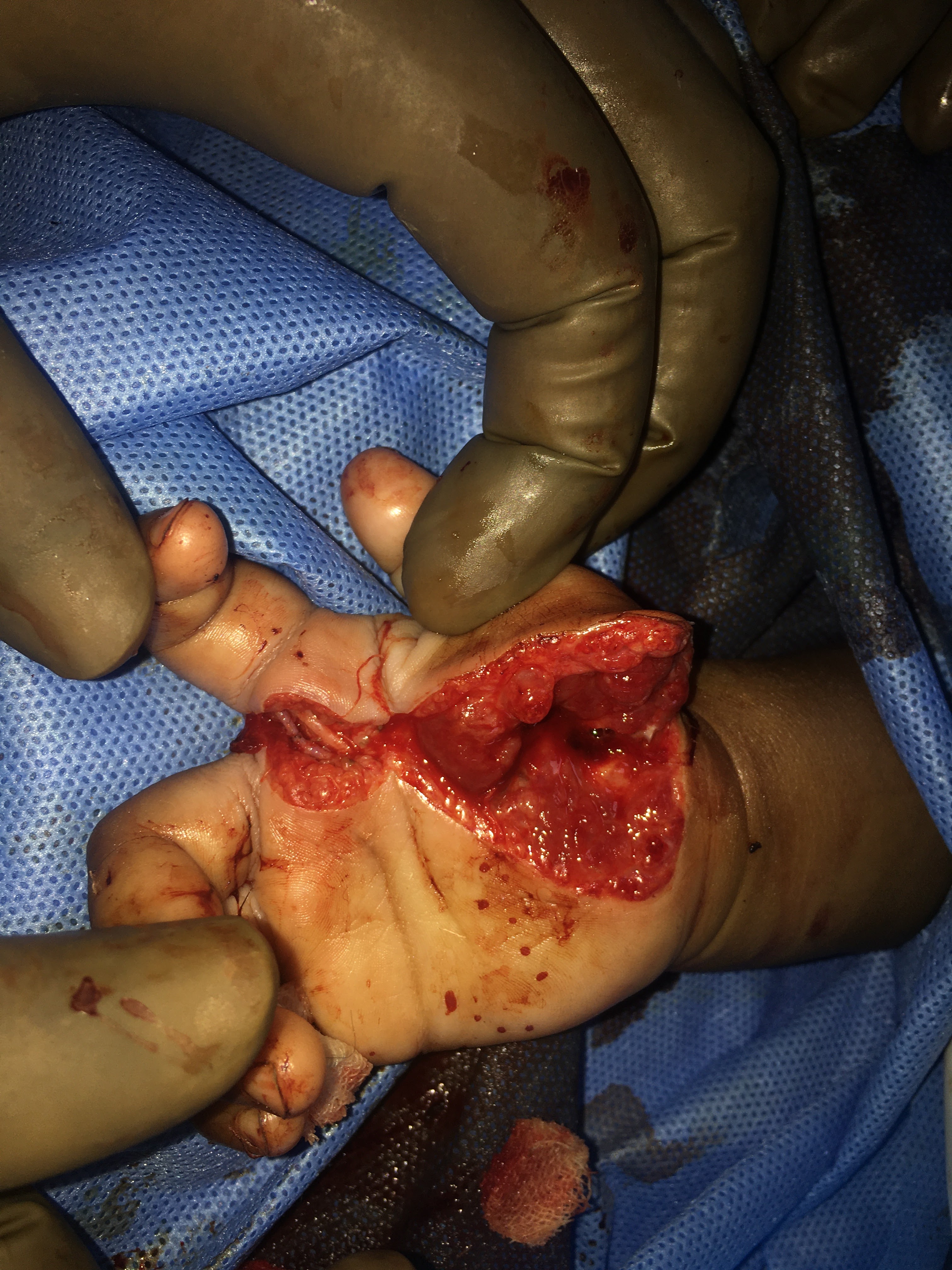
A patient presented with non healing ulcer in the buccal mucosa. On biopsy it was diagnosed as Squamous cell carcinoma and was involving the GB sulcus, cheek and also the overlying skin. Decision to do a composite resection of the tumor along with hemimandibulectomy was done. Earlier at the time of planning the reconstruction, the loss of cheek skin was envisaged and a double paddled(bipaddle) pectorals major musculocutaneous flap was planned. 
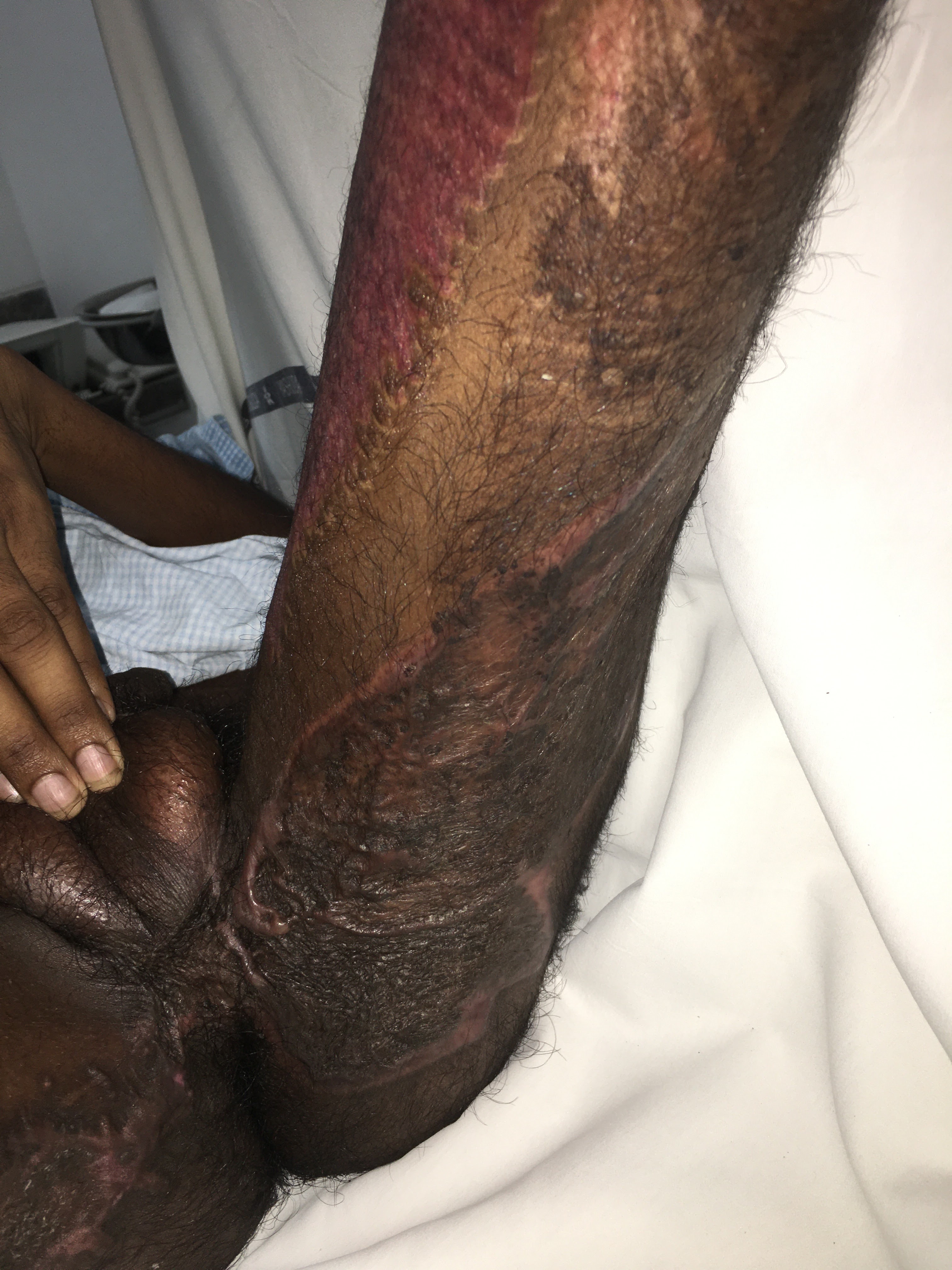
After the excision of the tumor the residual raw area is marked and the dimensions of the flap are marked. The pectorals major musculocutaneous flap with a long skin paddle was raised according to the standard technique with thin cuff of the muscle around the vascular pedicle of the flap. The flap is then tunneled through the subcutaneous space in to the defect. The intraoral part is inset first and the flap is turned to outside and flap is inset. The intervening segment at the angle of the mouth on the flap is deepithelialised and sutured to the angle of the mouth. 

Friday 19 June 2020

Latissimus dorsi musculocutaneous flap coverage for clavicular and supraclavicular defect after sarcoma excision


Soft tissue Sarcoma, over the clavicular and supraclavicular region. Treated with wide local excision and the residual raw area was covered with a Latissimus Dorsi Musculocutaneous flap, which was routed anteriorly into the defect. Post operatively flap is well settled in the defect and the healthy donor site with out any loss of skin. 

Upper eyelid, eyebrow, forehead and scalp Avulsion - Repair of the eyelid and rest of the wounds with restoration of normal appearance


Vikas Chapdi, met with a road traffic accident and sustained a severe injury to scalp , forehead, temporal and upper eyelid region on right side. 
Prompt repair of the upper eyelid , forehead, scalp and temporal region resulted in the good healing and very faint scars which are almost invisible. 

Crush injury of wrist and forearm with extensive degloving of the skin - Skin Grafting and Debridements


21 year old young Tanvir Fatima suffered a runover by a heavy vehicle over left hand, wrist and forearm. To her luck, there was no injury to the vascular supply of the hand, and it was only a degloving of the skin around the wrist and distal forearm. The devitalised skin was derided and negative pressure dressing was applied to improve the wound.
The final wound coverage was done by split thickness skin grafting. Complete functional rehabilitation of the hand and wrist apart from good take of the skin graft was achieved after 6 weeks after the final wound closure procedure. 

Microvascular repair of the Digital Artery of the Index finger in 7 month old child


Seven month old Harshita, accidentally sustained a cut injury over her palm over thenar eminence extending into the 2nd webspace damaging the ulnar digital artery of the index finger without damaging the digital nerve. The thenar eminence was cut damaging the muscles of the thenar eminence. 
the muscles of the thenar eminence were identified, isolated and repaired. 
Microvascular repair of the transected Digital artery of the index finger was done. 

Perineal and thing burns (deep) - Healed by Multiple Debridements and Split thickness Skin grafting


Muthyalu, a 25 year old worker in a ceramic factory, accidentally fell on a hot surface and suffered burns over his perineum, inner thighs and buttocks and legs and face. 
Most of the burns were full thickness deep grade 4 burns. Multiple debridements were done to excise the burn tissue. Simultaneously a Diverting colostomy was done to prevent soiling the burn wounds. A negative pressure wound dressing was applied to improve the wounds. Once the wounds were healthy without any infection, Wound coverage was done with split thickness skin grafting. 

Tuesday 17 March 2020

Extensive Anogenital warts - One and half year kid


Raw area post cauterization of the warts.


Focal recurrences after first cauterization 


Final photograph of healed area with minimal scarring


Severe Anogenital warts in 1 1/2 year old Patient from Somalia. Underwent treatment locally conservatively which was unsuccessful.  Due to microscopic presence of virus in the adjacent normal appearing skin, the chances of recurrence was explained to the parents. 
Electrocautery with ring tip was planned. 
Extensive electrocautery of the warts was done under general anesthesia. Cautery had to be run little deeper in the skin at some areas to cauterize the parts of warts which were deep. 
Post cautérisation the resultant raw area was managed by daily and regular dressing sometimes many times a day. After few weeks there was evidence of recurrent lesions on peri anal area and in the mucocutaneous junction of the anus and posterior commissure of vagina. Electrocautery was planned again and after few weeks post cauterization there was no evidence of new lesions. The raw areas post cauterization healed well with mild scarring. Patient was given instructions for focal chemical cauterization in case of focal recurrences in the form of imiquimod and the patient left for Somalia on 17-3-2020.